Human Anatomy Marieb Review Sheet Autonomic Nervous System
The nervous arrangement is the chief decision-making and communicating system of the torso. Every thought, action, and emotion reflects its activity. Its signaling device, or means of communicating with body cells, is electrical impulses, which are rapid and specific and cause near firsthand responses.
Functions of the Nervous System
To behave out its normal part, the nervous system has three overlapping functions.
- Monitoring changes. Much like a sentry, it uses its millions of sensory receptors to monitor changes occurring both inside and exterior the body; these changes are called stimuli, and the gathered information is chosen sensory input.
- Estimation of sensory input. It processes and interprets the sensory input and decides what should be done at each moment, a procedure called integration.
- Effects responses. It so effects a response by activating muscles or glands (effectors) via motor output.
- Mental action. The brain is the middle of mental activity, including consciousness, thinking, and retention.
- Homeostasis. This office depends on the ability of the nervous system to discover, interpret, and respond to changes in the internal and external weather condition. It tin help stimulate or inhibit the activities of other systems to assistance maintain a constant internal environment.
Beefcake of the Nervous System
The nervous organisation does non piece of work lone to regulate and maintain body homeostasis; the endocrine system is a second of import regulating system.
Organization of the Nervous System
We only have ane nervous organization, but, because of its complication, it is difficult to consider all of its parts at the same time; then, to simplify its report, nosotros separate information technology in terms of its structures (structural nomenclature) or in terms of its activities (functional nomenclature).
Structural Nomenclature
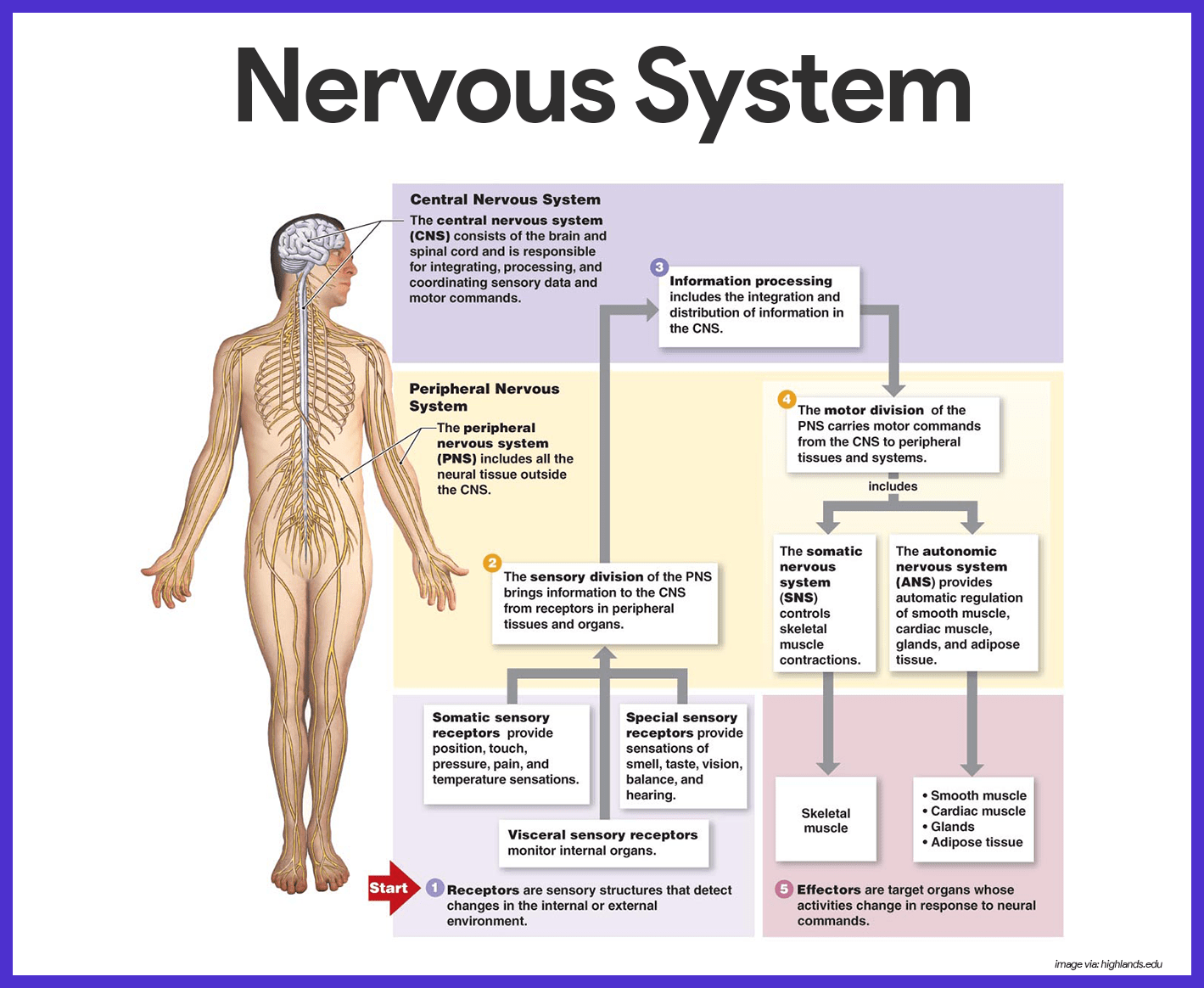
The structural classification, which includes all of the nervous system organs, has two subdivisions- the central nervous system and the peripheral nervous organization.
- Central nervous organisation (CNS). The CNS consists of the brain and spinal string, which occupy the dorsal body crenel and human activity as the integrating and command centers of the nervous organization
- Peripheral nervous system (PNS). The PNS, the part of the nervous organization outside the CNS, consists mainly of the fretfulness that extend from the brain and spinal cord.
Functional Classification
The functional classification scheme is concerned just with PNS structures.
- Sensory division. The sensory, or afferent division, consists of fretfulness (composed of nervus fibers) that convey impulses to the central nervous system from sensory receptors located in diverse parts of the torso.
- Somatic sensory fibers. Sensory fibers delivering impulses from the pare, skeletal muscles, and joints are called somatic sensory fibers.
- Visceral sensory fibers. Those that transmit impulses from the visceral organs are called visceral sensory fibers.
- Motor division. The motor, or efferent segmentation carries impulses from the CNS to effector organs, the muscles and glands; the motor division has 2 subdivisions: the somatic nervous organisation and the autonomic nervous system.
- Somatic nervous organisation. The somatic nervous system allows us to consciously, or voluntarily, control our skeletal muscles.
- Autonomic nervous system. The autonomic nervous system regulates events that are automated, or involuntary; this subdivision, normally called involuntary nervous arrangement, has two parts: the sympathetic and parasympathetic, which typically bring about opposite effects.
Nervous Tissue: Structure and Part
Fifty-fifty though it is complex, nervous tissue is fabricated up of just two principal types of cells- supporting cells and neurons.
Supporting Cells
Supporting cells in the CNS are "lumped together" as neuroglia, literally hateful "nervus mucilage".
- Neuroglia. Neuroglia include many types of cells that by and large support, insulate, and protect the delicate neurons; in improver, each of the dissimilar types of neuroglia, also only called either glia or glial cells,has special functions.
- Astrocytes. These are arable, star-shaped cells that account for nigh half of the neural tissue; astrocytes form a living barrier between the capillaries and neurons and play a role in making exchanges between the two so they could help protect neurons from harmful substances that might be in the blood.
- Microglia. These are spiderlike phagocytes that dispose of debris, including dead brain cells and bacteria.
- Ependymal cells. Ependymal cells are glial cells that line the central cavities of the brain and the spinal cord; the beating of their cilia helps to circulate the cerebrospinal fluid that fills those cavities and forms a protective absorber around the CNS.
- Oligodendrocytes. These are glia that wrap their flat extensions tightly around the nerve fibers, producing fat insulating coverings called myelin sheaths.
- Schwann cells. Schwann cells form the myelin sheaths effectually nervus fibers that are found in the PNS.
- Satellite cells. Satellite cells act every bit protective, cushioning cells.
Neurons
Neurons, also called nerve cells, are highly specialized to transmit messages (nerve impulses) from one role of the trunk to another.
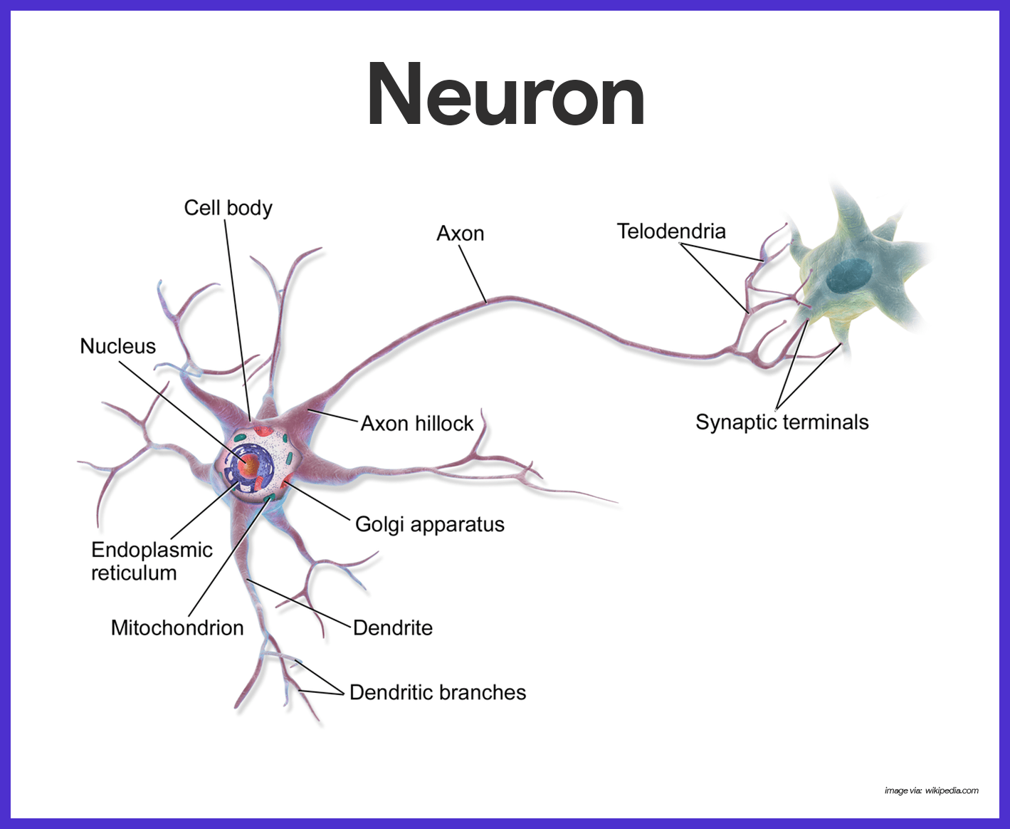
- Cell body. The jail cell body is the metabolic eye of the neuron; information technology has a transparent nucleus with a conspicuous nucleolus; the rough ER, called Nissl substance, and neurofibrils are especially abundant in the cell body.
- Processes. The armlike processes, or fibers, vary in length from microscopic to 3 to iv feet; dendrons convey incoming messages toward the cell trunk, while axons generate nerve impulses and typically conduct them away from the cell body.
- Axon hillock. Neurons may have hundreds of the branching dendrites, depending on the neuron type, only each neuron has just one axon, which arises from a conelike region of the cell body called the axon hillock.
- Axon terminals.These terminals incorporate hundreds of tiny vesicles, or bleary sacs that contain neurotransmitters.
- Synaptic crack. Each axon terminal is separated from the next neuron by a tiny gap called synaptic crack.
- Myelin sheaths. Most long nerve fibers are covered with a whitish, fat material called myelin, which has a waxy advent; myelin protects and insulates the fibers and increases the transmission charge per unit of nervus impulses.
- Nodes of Ranvier. Because the myelin sheath is formed by many private Schwann cells, information technology has gaps, or indentations, called nodes of Ranvier.
Nomenclature
Neurons may be classified either according to how they function or according to their structure.
- Functional classification. Functional classification groups neurons co-ordinate to the direction the nervus impulse is traveling relative to the CNS; on this basis, there are sensory, motor, and association neurons.
- Sensory neurons. Neurons carrying impulses from sensory receptors to the CNS are sensory, or afferent, neurons; sensory neurons keep united states of america informed about what is happening both inside and outside the torso.
- Motor neurons. Neurons carrying impulses from the CNS to the viscera and/or muscles and glands are motor, or efferent, neurons.
- Interneurons. The third category of neurons is known as the interneurons, or clan neurons; they connect the motor and sensory neurons in neural pathways.
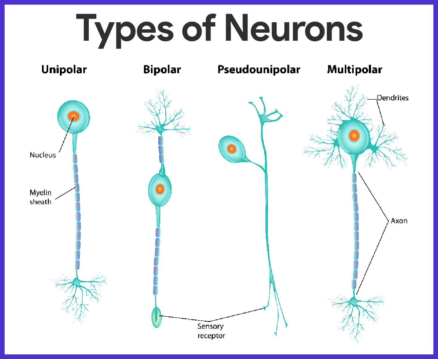
- Structural classification. Structural classification is based on the number of processes extending from the cell body.
- Multipolar neuron. If there are several processes, the neuron is a multipolar neuron; because all motor and association neurons are multipolar, this is the almost common structural type.
- Bipolar neurons. Neurons with two processes- an axon and a dendrite- are called bipolar neurons; these are rare in adults, found only in some special sense organs, where they act in sensory processing as receptor cells.
- Unipolar neurons. Unipolar neurons have a single process emerging from the cell's body, however, it is very short and divides almost immediately into proximal (central) and distal (peripheral) processes.
Central Nervous System
During embryonic development, the CNS beginning appears every bit a simple tube, the neural tube, which extends down the dorsal median plan of the developing embryo's body.
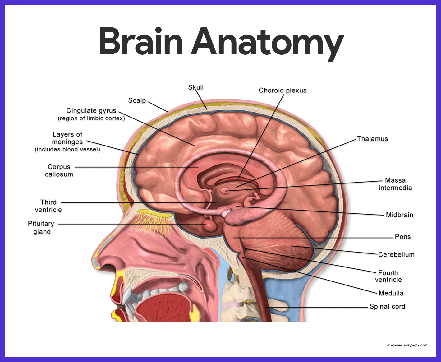
Encephalon
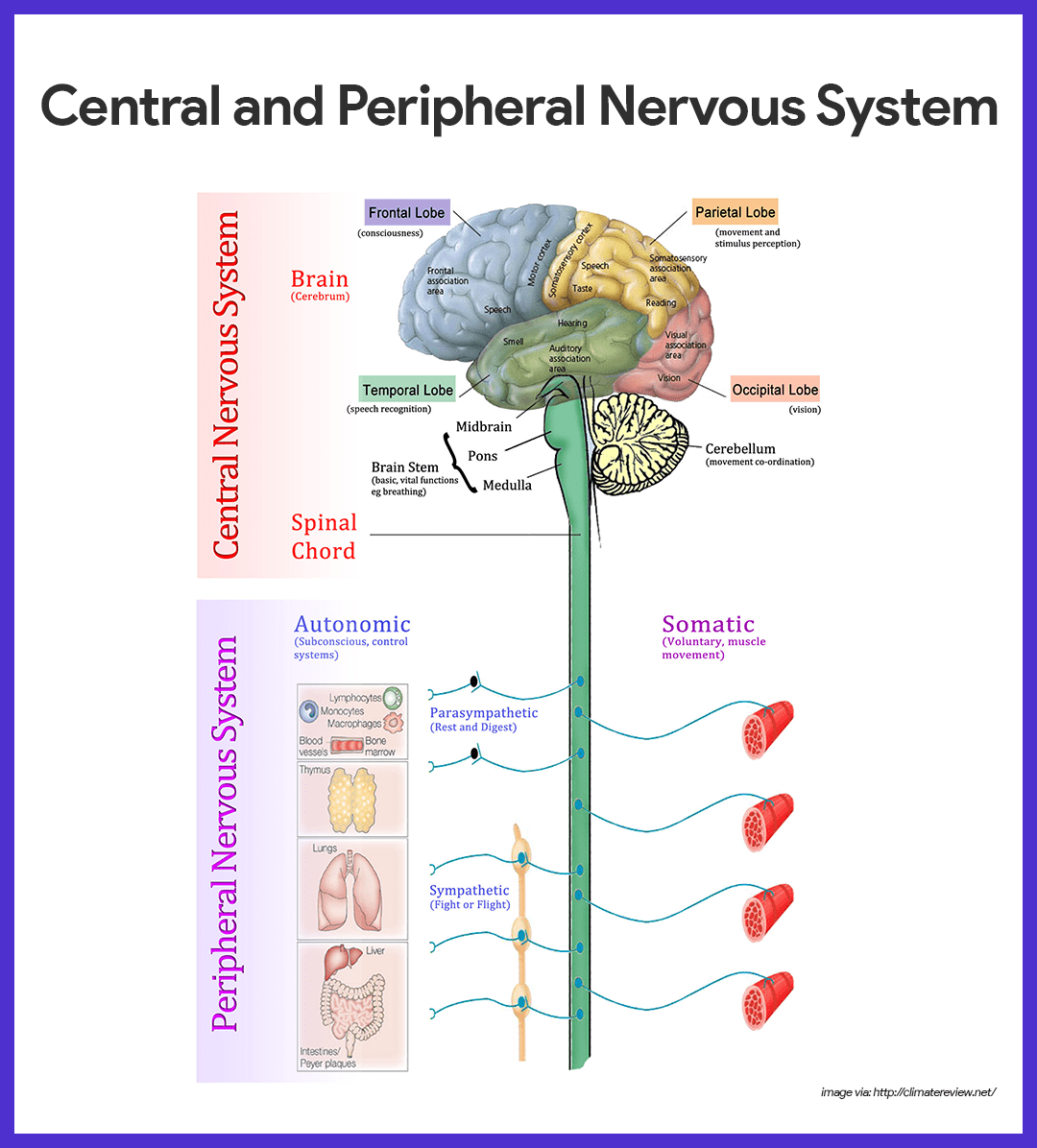
Because the encephalon is the largest and nigh complex mass of nervous tissue in the body, information technology is ordinarily discussed in terms of its 4 major regions – cerebral hemispheres, diencephalon, encephalon stem, and cerebellum.
Cerebral Hemispheres
The paired cognitive hemispheres, collectively called cerebrum, are the most superior function of the brain, and together are a good bargain larger than the other iii brain regions combined.
- Gyri. The entire surface of the cognitive hemispheres exhibits elevated ridges of tissue called gyri, separated past shallow grooves called sulci.
- Fissures. Less numerous are the deeper grooves of tissue called fissures, which separate large regions of the brain; the cognitive hemispheres are separated by a single deep fissure, the longitudinal fissure.
- Lobes. Other fissures or sulci dissever each hemisphere into a number of lobes, named for the cranial bones that prevarication over them.
- Regions of cognitive hemisphere. Each cerebral hemisphere has three basic regions: a superficial cortex of gray matter, an internal white matter, and the basal nuclei.
- Cerebral cortex. Oral communication, memory, logical and emotional response, likewise as consciousness, interpretation of sensation, and voluntary movement are all functions of neurons of the cerebral cortex.
- Parietal lobe. The primary somatic sensory surface area is located in the parietal lobe posterior to the central sulcus; impulses traveling from the body's sensory receptors are localized and interpreted in this area.
- Occipital lobe. The visual area is located in the posterior part of the occipital lobe.
- Temporal lobe. The auditory area is in the temporal lobe bordering the lateral sulcus, and the olfactory area is constitute deep inside the temporal lobe.
- Frontal lobe. The primary motor area, which allows united states of america to consciously move our skeletal muscles, is anterior to the central sulcus in the front end lobe.
- Pyramidal tract. The axons of these motor neurons form the major voluntary motor tract- the corticospinal or pyramidal tract, which descends to the cord.
- Broca's surface area. A specialized cortical surface area that is very involved in our ability to speak, Broca'south expanse, is constitute at the base of the precentral gyrus (the gyrus anterior to the key sulcus).
- Spoken language area. The speech surface area is located at the junction of the temporal, parietal, and occipital lobes; the spoken communication area allows one to audio out words.
- Cognitive white matter. The deeper cerebral white matter is compose of fiber tracts conveying impulses to, from, and within the cortex.
- Corpus callosum. 1 very large fiber tract, the corpus callosum, connect the cerbral hemispheres; such fiber tracts are called commisures.
- Fiber tracts. Association fiber tracts connect areas inside a hemisphere, and projection cobweb tracts connect the cerebrum with lower CNS centers.
- Basal nuclei. There are several islands of grey matter, chosen the basal nuclei, or basal ganglia, cached deep within the white matter of the cognitive hemispheres; information technology helps regulate the voluntary motor activities by modifying instructions sent to the skeletal muscles by the primary motor cortex.
Diencephalon
The diencephalon, or interbrain, sits atop the brain stem and is enclosed by the cerebral hemispheres.
- Thalamus. The thalamus, which encloses the shallow third ventricle of the brain, is a relay station for sensory impulses passing upward to the sensory cortex.
- Hypothalamus. The hypothalamus makes up the flooring of the diencephalon; information technology is an important autonomic nervous system eye because it plays a function in the regulation of trunk temperature, water rest, and metabolism; it is too the center for many drives and emotions, and every bit such, information technology is an important function of the and so-called limbic system or "emotional-visceral encephalon"; the hypothalamus as well regulates the pituitary gland and produces two hormones of its ain.
- Mammillary bodies. The mammillary bodies, reflex centers involved in olfaction (the sense of odor), bulge from the flooring of the hypothalamus posterior to the pituitary gland.
- Epithalamus. The epithalamus forms the roof of the tertiary ventricle; of import parts of the epithalamus are the pineal body (part of the endocrine system) and the choroid plexus of the third ventricle, which forms the cerebrospinal fluid.
Encephalon Stalk
The brain stem is nigh the size of a thumb in bore and approximately 3 inches long.
- Structures. Its structures are the midbrain, pons, and the medulla oblongata.
- Midbrain. The midbrain extends from the mammillary bodies to the pons inferiorly; it is composed of 2 jutting fiber tracts, the cerebral peduncles, which convey descending and ascending impulses.
- Corpora quadrigemina. Dorsally located are four rounded protrusions called the corpora quadrigemina because they remind some anatomist of two pairs of twins; these jutting nuclei are reflex centers involved in vision and hearing.
- Pons. The pons is a rounded structure that protrudes but beneath the midbrain, and this area of the brain stem is mostly cobweb tracts; nonetheless, it does have important nuclei involved in the control of breathing.
- Medulla oblongata. The medulla oblongata is the most inferior part of the brain stem; it contains nuclei that regulate vital visceral activities; it contains centers that control heart rate, blood pressure, animate, swallowing, and airsickness amid others.
- Reticular formation. Extending the entire length of the encephalon stem is a lengthened mass of gray matter, the reticular formation; the neurons of the reticular formation are involved in motor command of the visceral organs; a special grouping of reticular formation neurons, the reticular activating system (RAS), plays a office in consciousness and the awake/sleep cycles.
Cerebellum
The large, cauliflower-like cerebellum projects dorsally from nether the occipital lobe of the cerebrum.
- Structure. Like the cerebrum. the cerebellum has two hemispheres and a convoluted surface; information technology besides has an outer cortex made upward of gray thing and an inner region of white matter.
- Function. The cerebellum provides precise timing for skeletal musculus action and controls our residue and equilibrium.
- Coverage. Fibers reach the cerebellum from the equilibrium apparatus of the inner ear, the eye, the proprioceptors of the skeletal muscles and tendons, and many other areas.
Protection of the Primal Nervous System
Nervous tissue is very soft and delicate, and the irreplaceable neurons are injured by even the slightest force per unit area, so nature has tried to protect the brain and the spinal cord past enclosing them inside os (the skull and vertebral column), membranes (the meninges), and a watery absorber (cerebrospinal fluid).
Meninges
The three connective tissue membranes roofing and protecting the CNS structures are the meninges.
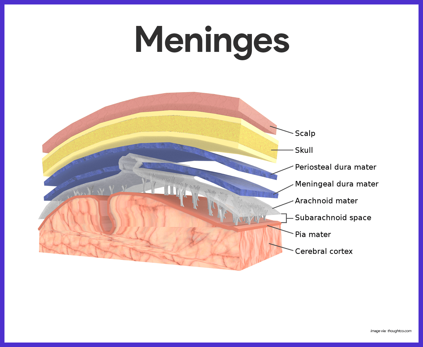
- Dura mater. The outermost layer, the leathery dura mater, is a double layered membrane where it surrounds the brain; one of its layer is attached to the inner surface of the skull, forming the periosteum (periosteal layer); the other, called the meningeal layer, forms the outermost roofing of the brain and continues every bit the dura mater of the spinal cord.
- Falx cerebri.In several places, the inner dural membrane extends in to course a fold that attaches the encephalon to the cranial crenel, and 1 of these folds is the falx cerebri.
- Tentorium cerebelli. The tentorium cereberi separates the cerebellum from the cerebrum.
- Arachnoid mater. The middle layer is the weblike arachnoid mater; its threadlike extensions span the subarachnoid space to attach it to the innermost membrane.
- Pia mater. The delicate pia mater, the innermost meningeal layer, clings tightly to the surface of the brain and spinal string, following every fold.
Cerebrospinal Fluid
Cerebrospinal fluid (CSF) is a watery "broth" like in its makeup to blood plasma, from which it forms.
- Contents. The CSF contains less protein and more vitamin C, and glucose.
- Choroid plexus. CSF is continually formed from claret by the choroid plexuses; choroid plexuses are clusters of capillaries hanging from the "roof" in each of the brain's ventricles.
- Function. The CSF in and around the brain and cord forms a watery cushion that protects the delicate nervous tissue from blows and other trauma.
- Normal volume. CSF forms and drains at a constant rate so that its normal pressure level and volume (150 ml-about half a loving cup) are maintained.
- Lumbar tap. The CSF sample for testing is obtained past a process chosen lumbar or spinal tap;because the withdrawal of fluid for testing decreases CSF fluid pressure, the patient must remain in a horizontal position (lying downward) for 6 to 12 hours afterward the procedure to foreclose an agonizingly painful "spinal headache".
The Blood-Encephalon Barrier
No other body organ is so absolutely dependent on a constant internal environment as is the brain, and so the blood-brain barrier is there to protect it.
- Function. The neurons are kept separated from bloodborne substances past the so-chosen blood-encephalon bulwark, equanimous of the least permeable capillaries in the whole body.
- Substances allowed. Of water-soluble substances, only h2o, glucose, and essential amino acids pass easily through the walls of these capillaries.
- Prohibited substances. Metabolic wastes, such every bit toxins, urea, proteins, and most drugs are prevented from entering the brain tissue.
- Fat-soluble substances. The blood-brain barrier is virtually useless confronting fats, respiratory gases, and other fat-soluble molecules that diffuse easily through all plasma membranes.
Spinal String
The cylindrical spinal cord is a glistening white continuation of the brain stem.

- Length. The spinal string is approximately 17 inches (42 cm) long.
- Major part. The spinal cord provides a 2-style conduction pathway to and from the brain, and it is a major reflex center (spinal reflexes are completed at this level).
- Location. Enclosed within the vertebral column, the spinal cord extends from the foramen magnum of the skull to the outset or 2nd lumbar vertebra, where it ends just below the ribs.
- Meninges. Similar the brain, the spinal string is cushioned and protected past the meninges; meningeal coverings do not end at the second lumbar vertebra but instead extend well beyond the end of the spinal cord in the vertebral culvert.
- Spinal nerves. In humans, 31 pairs of spinal nerves arise from the cord and exit from the vertebral cavalcade to serve the body area shut by.
- Cauda equina. The collection of spinal nerves at the inferior end of the vertebral canal is chosen cauda equina because information technology looks and so much like a horse's tail.
Gray Matter of the Spinal String and Spinal Roots
The gray matter of the spinal string looks like a butterfly or a letter H in cross department.
- Projections. The two posterior projections are the dorsal, or posterior, horns; the two anterior projections are the ventral, or anterior, horns.
- Cardinal culvert. The gray matter surrounds the cardinal canal of the string, which contains CSF.
- Dorsal root ganglion. The cell bodies of sensory neurons, whose fibers enter the cord by the dorsal root, are institute in an enlarged expanse called dorsal root ganglion; if the dorsal root or its ganglion is damaged, sensation from the body area served will be lost.
- Dorsal horns. The dorsal horns contain interneurons.
- Ventral horns. The ventral horns of gray thing contain cell bodies of motor neurons of the somatic nervous system, which ship their axons out the ventral root of the cord.
- Spinal nerves. The dorsal and ventral roots fuse to grade the spinal nerves.
White Matter of the Spinal Cord
White matter of the spinal cord is equanimous of myelinated cobweb tracts- some running to higher centers, some traveling from the brain to the string, and some conducting impulses from one side of the spinal string to the other.
- Regions. Considering of the irregular shape of the gray thing, the white matter on each side of the cord is divided into three regions- the dorsal, lateral, and ventral columns; each of the columns contains a number of fiber tracts made up of axon with the same destination and role.
- Sensory tracts. Tracts conducting sensory impulses to the brain are sensory, or afferent, tracts.
- Motor tracts. Those carrying impulses from the brain to skeletal muscles are motor, or efferent, tracts.
Peripheral Nervous System
The peripheral nervous system consists of nerves and scattered groups of neuronal jail cell bodies (ganglia) found outside the CNS.
Structure of a Nerve
A nerve is a bundle of neuron fibers constitute outside the CNS.
- Endoneurium. Each fiber is surrounded past a fragile connective tissue sheath, an endoneurium.
- Perimeurium. Groups of fibers are bound by a coarser connective tissue wrapping, the perineurium, to course cobweb bundles, or fascicles.
- Epineurium. Finally, all the fascicles are bound together by a tough fibrous sheath, the epineurium, to course the cordlike nerve.
- Mixed nerves. Nerves carrying both sensory and motor fibers are chosen mixed nerves.
- Sensory nerves. Fretfulness that carry impulses toward the CNS only are called sensory, or afferent, nerves.
- Motor nerves. Those that carry only motor fibers are motor, or efferent, nerves.
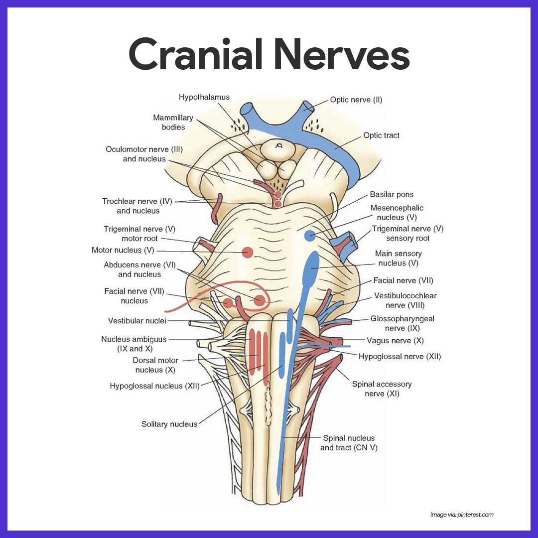
Cranial Fretfulness
The 12 pairs of cranial nerves primarily serve the head and the cervix.
- Olfactory. Fibers arise from the olfactory receptors in the nasal mucosa and synapse with the olfactory bulbs; its function is purely sensory, and information technology carries impulses for the sense of smell.
- Optic. Fibers arise from the retina of the center and form the optic nerve; its role is purely sensory, and carries impulses for vision.
- Oculomotor. Fibers run from the midbrain to the eye; it supplies motor fibers to four of the six muscles (superior, inferior, and medial rectus, and inferior oblique) that direct the eyeball; to the eyelid; and to the internal eye muscles controlling lens shape and pupil size.
- Trochlear. Fibers run from the midbrain to the centre; it supplies motor fibers for ane external eye muscle ( superior oblique).
- Trigeminal. Fibers emerge from the pons and class iii divisions that run to the confront; it conducts sensory impulses from the skin of the face up and mucosa of the nose and rima oris; also contains motor fibers that activate the chewing muscles.
- Abducens. Fibers get out the pons and run to the eye; it supplies motor fibers to the lateral rectus muscle, which rolls the centre laterally.
- Facial. Fibers leave the pons and run to the face up; it activates the muscles of facial expression and the lacrimal and salivary glands; carries sensory impulses from the taste buds of the inductive tongue.
- Vestibulocochlear. fibers run from the equilibrium and hearing receptors of the inner ear to the brain stem; its function is purely sensory; vestibular branch transmits impulses for the sense of residual, and cochlear co-operative transmits impulses for the sense of hearing.
- Glossopharyngeal. Fibers emerge from the medulla and run to the pharynx; it supplies motor fibers to the pharynx (throat) that promote swallowing and saliva production; it carries sensory impulses from the taste buds of the posterior natural language and from pressure level receptors of the carotid artery.
- Vagus. Fibers emerge from the medulla and descend into the thorax and intestinal crenel; the fibers carry sensory impulses from and motor impulses to the throat, larynx, and the abdominal and thoracic viscera; nigh motor fibers are parasympathetic fibers that promote digestive activeness and assist regulate center activity.
- Accompaniment. Fiber arise from the medulla and superior spinal cord and travel to muscles of the neck and dorsum; generally motor fiber that activate the sternocleidomastoid and trapezius muscles.
- Hypoglossal. Fibers run from the medulla to the tongue; motor fibers control natural language movements;; sensory fibers conduct impulses from the tongue.
Spinal Nerves and Nerve Plexuses
The 31 pairs of human spinal nerves are formed by the combination of the ventral and dorsal roots of the spinal string.
- Rami. Almost immediately subsequently being formed, each spinal nerve divides into dorsal and ventral rami, making each spinal nerve merely about 1/two inch long; the rami contains both sensory and motor fibers.
- Dorsal rami. The smaller dorsal rami serve the peel and muscles of the posterior body body.
- Ventral rami. The ventral rami of spinal nerves T1 through T12 form the intercostal nerves, which supply the muscles between the ribs and the skin and muscles of the anterior and lateral trunk.
- Cervical plexus. The cervical plexus originates from the C1-C5, and phrenic nerve is an important nerve; it serves the diaphragm, and skin and muscles of the shoulder and neck.
- Brachial plexus. The axillary nerve serve the deltoid muscles and skin of the shoulder, muscles, and skin of superior thorax; the radial nerve serves the triceps and extensor muscles of the forearm, and the pare of the posterior upper limb; the median nerve serves the flexor muscles and skin of the forearm and some muscles of the hand; the musculocutaneous nerve serves the flexor muscles of arm and the pare of the lateral forearm; and the ulnar nerve serves some flexor muscles of forearm; wrist and many hand muscles, and the skin of the manus.
- Lumbar plexus. The femoral nerve serves the lower abdomen, anterior and medial thigh muscles, and the pare of the anteromedial leg and thigh; the obturator nervus serves the adductor muscles of the medial thigh and small hip muscles, and the pare of the medial thigh and hip joint.
- Sacral plexus. The sciatic nerve (largest nervus in the body) serves the lower torso and posterior surface of the thigh, and it splits into the common fibular and tibial nerves; the mutual fibular nerve serves the lateral aspect of the leg and pes, while the tibial nerve serves the posterior aspect of leg and foot; the superior and inferior gluteal fretfulness serve the gluteal muscles of the hip.
Autonomic Nervous System
The autonomic nervous system (ANS) is the motor subdivision of the PNS that controls torso activities automatically.
- Composition. It is composed of a specialized group of neurons that regulate cardiac muscle, smooth muscles, and glands.
- Function. At every moment, signals inundation from the visceral organs into the CNS, and the automatic nerves make adjustments every bit necessary to all-time support body activities.
- Divisions. The ANS has ii arms: the sympathetic partition and the parasympathetic partitioning.
Anatomy of the Parasympathetic Division
The parasympathetic partition allows us to "unwind" and conserve energy.
- Preganglionic neurons. The preganglionic neurons of the parasympathetic partitioning are located in brain nuclei of several cranial nerves- III, VII, Ix, and Ten (the vagus beingness the most important of these) and in the S2 through S4 levels of the spinal cord.
- Craniosacral division. The parasympathetic division is likewise called the craniosacral division; the neurons of the cranial region transport their axons out in cranial nerves to serve the head and neck organs.
- Pelvic splanchnic nerves. In the sacral region, the preganglionic axons leave the spinal cord and form the pelvic splanchnic fretfulness, also chosen the pelvic nerves, which travel to the pelvic crenel.
Anatomy of the Sympathetic Partitioning
The sympathetic sectionalization mobilizes the body during extreme situations, and is also called the thoracolumbar partition considering its preganglionic neurons are in the greyness thing of the spinal cord from T1 through L2.
- Ramus communicans. The preganglionic axons exit the cord in the ventral root, enter the spinal nervus, and then pass through a ramus communicans, or small communicating branch, to enter a sympathetic concatenation ganglion.
- Sympathetic chain. The sympathetic trunk, or chain, lies along the vertebral column on each side.
- Splanchnic nerves. After information technology reaches the ganglion, the axon may synapse with the second neuron in the sympathetic chain at the same or a different level, or the axon may through the ganglion without synapsing and form role of the splanchnic nerves.
- Collateral ganglion. The splanchnic nerves travel to the viscera to synapse with the ganglionic neuron, found in a collateral ganglion anterior to the vertebral column.
Physiology of the Nervous Arrangement
The physiology of the nervous system involves a complex journey of impulses.
Nerve Impulse
Neurons take two major functional properties: irritability, the power to answer to a stimulus and catechumen it into a nerve impulse, and conductivity, the ability to transmit the impulse to other neurons, muscles, or glands.
- Electrical conditions of a resting neuron's membrane. The plasma membrane of a resting, or inactive, neuron is polarized, which means that in that location are fewer positive ions sitting on the inner face of the neuron'southward plasma membrane than there are on its outer surface; every bit long as the inside remains more negative than the outside, the neuron will stay inactive.
- Action potential initiation and generation. Most neuron in the trunk are excited by neurotransmitters released by other neurons; regardless what the stimulus is, the result is ever the same- the permeability properties of the cell'southward plasma membrane change for a very brief period.
- Depolarization. The inward rush of sodium ions changes the polarity of the neuron's membrane at that site, an consequence called depolarization.
- Graded potential. Locally, the inside is now more positive, and the outside is less positive, a situation chosen graded potential.
- Nervus impulse. If the stimulus is stiff enough, the local depolarization activates the neuron to initiate and transmit a long-distance bespeak called action potential, also called a nerve impulse; the nerve impulse is an all-or-none response; it is either propagated over the entire axon, or it doesn't happen at all;it never goes partway along an axon'due south length, nor does information technology die out with altitude every bit exercise graded potential.
- Repolarization. The outflow of positive ions from the prison cell restores the electrical weather at the membrane to the polarized or resting, state, an event called repolarization; until a repolarization occurs, a neuron cannot conduct another impulse.
- Saltatory conduction. Fibers that have myelin sheaths conduct impulses much faster because the nerve impulse literally jumps, or leaps, from node to node along the length of the cobweb; this occurs considering no electrical current tin can flow across the axon membrane where there is fatty myelin insulation.
The Nervus Impulse Pathway
How the nerve impulse actually works is detailed below.
- Resting membrane electrical conditions. The external confront of the membrane is slightly positive; its internal face is slightly negative; the chief extracellular ion is sodium, whereas the chief intracellular ion is potassium; the membrane is relatively permeable to both ions.
- Stimulus initiates local depolarization. A stimulus changes the permeability of a "patch" of the membrane, and sodium ions diffuse rapidly into the cell; this changes the polarity of the membrane (the within becomes more positive; the outside becomes more negative) at that site.
- Depolarization and generation of an activity potential. If the stimulus is strong enough, depolarization causes membrane polarity to be completely reversed and an activity potential is initiated.
- Propagation of the activeness potential. Depolarization of the starting time membrane patch causes permeability changes in the side by side membrane, and the events described in (b) are repeated; thus, the action potential propagates quickly along the unabridged length of the membrane.
- Repolarization. Potassium ions diffuse out of the cell as the membrane permeability changes once more, restoring the negative charge on the inside of the membrane and the positive charge on the outside surface; repolarization occurs in the same direction equally depolarization.
Communication of Neurons at Synapses
The events occurring at the synapse are arranged below.
- Arrival. The activity potential arrives at the axon terminal.
- Fusion. The vesicle fuses with plasma membrane.
- Release. Neurotransmitter is released into synaptic cleft.
- Bounden. Neurotransmitter binds to receptor on receiving neuron's cease.
- Opening. The ion aqueduct opens.
- Closing. Once the neurotransmitter is broken downwardly and released, the ion channel shut.
Autonomic Operation
Trunk organs served by the autonomic nervous organisation receive fibers from both divisions.
- Antagonistic upshot. When both divisions serve the same organ, they cause antagonistic furnishings, mainly considering their post ganglionic axons release different transmitters.
- Cholinergic fibers. The parasympathetic fibers called cholinergic fibers, release acetylcholine.
- Adrenergic fibers. The sympathetic postganglionic fibers, called adrenergic fibers, release norepinephrine.
- Preganglionic axons. The preganglionic axons of both divisions release acetylcholine.
Sympathetic Division
The sympathetic partition is often referred to every bit the "fight-or-flight" system.
- Signs of sympathetic nervous system activities. A pounding heart; rapid, deep animate; cold, sweaty peel; a prickly scalp, and dilated pupils are certain signs sympathetic nervous system activities.
- Effects. Under such weather, the sympathetic nervous system increases heart rate, blood pressure, and blood glucose levels; dilates the bronchioles of the lungs; and brings about many other effects that aid the individual cope with the stressor.
- Duration of the outcome. The effects of sympathetic nervous system activation continue for several minutes until its hormones are destroyed by the liver.
- Part. Its role is to provide the best weather condition for responding to some threat, whether the all-time response is to run, to see better, or to think more conspicuously.
Parasympathetic Sectionalisation
The parasympathetic partitioning is almost active when the torso is at rest and not threatened in any way.
- Part. This division, sometimes chosen the "resting-and-digesting" system, is chiefly concerned with promoting normal digestion, with elimination of feces and urine, and with conserving torso free energy, particularly by decreasing demands on the cardiovascular system.
- Relaxed country. Claret pressure and centre and respiratory rates charge per unit being regulated at normal levels, the digestive tract is actively digesting food, and the skin is warm (indicating that there is no demand to divert blood to skeletal muscles or vital organs.
- Optical land. The eye pupils are constricted to protect the retinas from excessive dissentious light, and the lenses of the eye are "set" for shut vision.
Practice Quiz: Nervous System Anatomy and Physiology
Here's a ten-item quiz about the report guide. Delight visit our nursing test banking company page for more NCLEX practise questions.
1.The cell trunk of all sensory neurons is located within the:
A. Dorsal gray horn
B. Dorsal root ganglion
C. Spinal cord
D. Encephalon
1. Answer: B. Dorsal root ganglion
- B: The jail cell bodies of the sensory neurons leading to the spinal cord are located in clusters, the dorsal root ganglia (DRG), adjacent to the spinal cord.
- A: Dorsal gray horn is a mass of gray matter plant in every segment of the spinal string and is responsible for the processing arrangement of sensory signals that travel within the spinal cord,
- C: The spinal string is a long, thin, tubular bundle of nervous tissue and support cells that extends from the foramen magnum at the base of operations of the skull to the second lumbar vertebrae.
- D: Brain serves equally a command middle for the human nervous organisation. It receives input from the sensory organs and sends output to the muscles.
2. Which of the following is referred to as either physical barriers or physiologic processes (ship organisation) that carve up the circulating claret from the brain extracellular fluid in the cardinal nervous organization (CNS)?
A. Circle of Willis
B. Blood-brain bulwark
C. Corticobulbar projections
D. Lateral corticospinal tract
2. Answer: B. Blood-encephalon barrier
- B: Blood-brain barrier serves to restrict and control the motion of substances betwixt the general circulation and brain extracellular fluid.
- A:The Circumvolve of Willis is an arterial polygon formed as the internal carotid and vertebral systems anastomose around the optic chiasm or chiasma (partial crossing of the optic nerve. It provides blood to the encephalon and neighboring structures.
- C: Corticobulbar projections take several functions including voluntary control over cranial nerves, relay to the cerebellum, activation of other descending pathways and modulation of sensory processing.
- D: Lateral corticospinal tract are responsible for controlling the speed and precision of skilled movements of the hands
3. A male client was involved in a vehicular blow and developed an amnesia. He is likely having a damaged in which of the following?
A. Hypothalamus
B. Thalamus
C. Cerebrum
D. Hippocampus
three. Answer: D. Hippocampus
- D: The hippocampus is associated mainly with memory, especially, long-term retentivity.
- A:Hypothalamus controls vital actual functions such as hunger, thirst, body temperature, and hormone secretion.
- B: Thalamusinfluences mood and registers an unlocalized, uncomfortable perception of hurting.
- C:Cerebrumcontrols encephalon functions such as language, logic, reasoning, and creativity.
four. A client went to the emergency department with a possible brain damage as evidenced by loss of coordination of motor motility, and staggering, wide based walking. The client is nearly probable having a impairment in the:
A. Medulla Oblongata
B. Cerebrum
C. Pons
D. Cerebellum
iv. Answer: D. Cerebellum
- D: The cerebellum is involved in balance, maintenance of muscle tone, and coordination of fine motor motility.
- A:Breathing, vomiting, sneezing, coughing, and swallowing reflexes are coordinated in the medulla oblongata.
- B: Cerebrum controls encephalon functions such as linguistic communication, logic, reasoning, and creativity.
- C:Pons serves as a message station betwixt several areas of the encephalon. It helps relay messages from the cortex and the cerebellum.
five. It is a blazon of nerve cell that protects CNS from infection and become phagocytic in response to inflammation
A. Schwann cells
B. Ependymal cells
C. Microglia
D. Astrocytes
5. Answer: C. Microglia
- C:Microglia helps remove bacteria and cell debris from the CNS.
- A: Schwann cells grade myelin sheaths effectually axons, or enclose unmyelinated axons in the peripheral nervous system.
- B: Ependymal cells line ventricles of the encephalon, circulate cerebrospinal fluid; some grade choroid plexuses, which produce CSF.
- D: Astrocytes serves every bit the major supporting tissue in the CNS and contribute to the blood-brain barrier.
half dozen. Which of the following manifestations is consequent with a client who sustained a traumatic left parietal lobe injury?
A. Difficulty with writing, inability to perceive objects usually
B. Short term memory, blurred vision
C. Altered personality and affective behavior
D. Loss of fine movements and strength of the arms, hands, and fingers
6. Respond: A. Difficulty with writing, inability to perceive objects unremarkably
- A: Damage to the left parietal lobe can result in what is called Gerstmann's Syndrome which is characterized past difficulty with writing (agraphia), difficulty with mathematics (acalculia), and the inability to perceive objects ordinarily (agnosia).
- B & C: These are symptoms of a temporal lobe damage.
- D: Disturbance of such motor office is related to a frontal lobe harm.
7. Which among cranial nerves are mixed nerves that supply parasympathetic to viscera of thorax and abdomen?
A. Vagus nerves
B. Trigeminal nerves
C. Accessory nerves
D. Abducens nerves
7. Answer: A. Vagus nerves
- A:Vagus nerves provides sensory fibers that carry impulses from the lining of the larynx, pharynx, esophagus, thorax and abdomen to the encephalon.
- B: Trigeminal nerves provide sensory impulses to face up and teeth; motor to muscles of mastication.
- C :Accessory nerves carries impulses to muscles of the soft palate, throat, and larynx. The spinal branches pass down into the neck and back.
- D: Abducens nerves carry motor impulses to one extrinsic eye muscle.
eight. The ___________ is the innermost meningeal layer, clings tightly to the surface of the brain and spinal cord, following every fold.
A. Arachnoid mater
B. Pia mater
C. Dura mater
D. Tentorium cerebelli
8. Respond: B. Pia mater
- B: The innermost layer that contours closely to the many folds and crevices of the brain is called the pia mater.
- A: The arachnoid or arachnoid mater is the center layer of the meninges.
- C: Dura mater is the outermost layer of the meninges.
- D: Tentorium cerebelli is an extension of the dura mater that separates the cerebellum from the junior portion of the occipital lobes.
9. Nurse Jerick is performing a caloric testing of the vestibulo-ocular reflex of an unconscious client. Warm water is poured into the left auditory canal. The customer exhibit cohabit eye movement toward the right followed by nystagmus toward the left. The nurse understands that the customer is exhibiting a:
A. Midbrain lesion
B. Coma.
C. Intact brainstem
E. Gaze paralysis
nine. Respond: C. Intact brainstem
- C: A caloric testing of using warm water in a client with an intact encephalon stem: Pouring of warm h2o into the left ear leads to heart motility to the right and a direction of the nystagmus is on the left (aforementioned side).
10. Which of the following is the rationale to perform a spinal tap on a newly diagnosed customer with leukemia?
A. To dominion out bacterial meningitis
B. To cheque if leukemia has spread to the cerebral spinal fluid
C. To allocate the blazon of leukemia
D. To lower the pressure level in the brain
10. Respond: B. To check if leukemia has spread to the cognitive spinal fluid
- B: A spinal tap tin determine if leukemia has infiltrated to the cerebral spinal fluid (CSF).
- A: A spinal tap can be washed to dominion out bacterial meningitis merely this is not the reason for the exam on a customer with leukemia.
- C & D: Spinal tap is non done to identify the classification of leukemia and to decrease intracranial pressure.
Source: https://nurseslabs.com/nervous-system/
0 Response to "Human Anatomy Marieb Review Sheet Autonomic Nervous System"
Post a Comment